how do they x ray babies hips
Generally the benefits of the diagnostic information from an X-ray outweigh the potential risk to a baby. Its a cast that goes around both hips and down the leg to keep the hips aligned.

Pin On Veterinary Medicine
The age groups were based on exposures suitable for tissue thickness in the direction of the X-ray beam of a patient of averagestandard size in that age group for each projection.

. Appointments and Referrals. The X-ray image is black and white. You will go in the room with him he will need to be stripped from the waist down they will take x-rays of him flat on his back legs dead straight and together you wil be able to hold him in this position then an x-ray of his still on his back with his knees bent facing outwards and the soles of his.
Totaleclipse 07092007 1731. They give your healthcare provider information about structures inside the body. They do this by gently pushing and pulling the babys thigh bones to see if they are loose in the hip socket.
For some reason the left hip is said to be more frequently affected 4. The black-and-white images show the internal structures of the hip including the ball-shaped top of the thighbone femoral head and its socket acetabulum in the pelvic bone. The hip ultrasound will show the healthcare provider the position and shape of the hip joint.
They can penetrate your body. But for babies with an abnormal physical exam or major risk factors for developmental dysplasia of the hip or DDH family history Breech position etc the AAP supports referral for. Dense body parts such as bones block the passage of the X-ray beam.
They are of more concern. X-rays can be taken once your baby is 3 months old. You would have to x-ray your arm or leg more than 5000 times in order to reach 5 rad of exposure to your unborn baby.
X-rays are forms of radiant energy like light or radio waves. If she does have it they may try to brace it first. An ultrasound machine sends sound waves into the hip area and images are recorded on a computer.
Because they spin around the body taking multiple images CT scans can deliver radiation doses that are up to 200 times higher than an. When should I order an X-ray rather than an ultrasound to diagnose a musculoskeletal problem in an infant. How Do They Xray Babies Head.
What Are the. 812 years average 10 years. The International Hip Dysplasia Institute offers the following advice for choosing baby carriers photo source.
51-370 millirad for x-rays of the hip and femur thighbone It is very rare for a single diagnostic x-ray to exceed even 5 rad. 200-245 millirad for an x-ray of the abdomen. It is put on by an orthopedic surgeon while using.
Around 6 months of age enough bone is present in an infant hip to make an X-ray more accurate than ultrasound. 37 years average 5 years. However if you received a large number of abdominal X-rays over a short period before you were aware of your pregnancy your baby could be affected.
However x-rays of the mothers lower torso - abdomen stomach pelvis lower back or kidneys - may expose the unborn child to the direct x-ray beam. The scan can be done on babies up to about 6 months of age. In some institutions pediatric patients with hip pain may be assessed with a single frog leg view to reduce radiation exposure.
The American Academy of Pediatrics does not recommend routine ultrasounds for every infant. X-rays have more energy than rays of visible light or radio waves. The purpose of this study is to report US results and follow-up of.
The doctor first checks your babys hips in the hospital after birth. About one in eight scans ordered for kids is a CT scan. Because of the risk of developmental dysplasia of the hip in infants born breech-despite a normal physical exam-the American Academy of Pediatrics AAP guidelines recommend ultrasound US hip imaging at 6 weeks of age for breech females and optional imaging for breech males.
The main idea is to choose a sling that supports the legs taking the pressure off of the hips. If the pediatric patient can be kept still using other methods such as distraction techniques or swaddling this is ideal to avoid scattered radiation to parents and staff 3. The possibility of an X-ray during pregnancy causing harm to your unborn child is very small.
In fact it seems that proper baby wearing can help promote proper development of the babys hips. An X-ray is a safe and painless test that uses a small amount of radiation to make an image of bones organs and other parts of the body. 1317 years average 15 years.
X-rays are a kind of imaging test. These tests expose children to low doses of radiation. 40-240 millirad for an x-ray of the pelvis.
Radiation protection considerations. If it persists they may put on a spica cast.

Pin By Meg Carter On Ortho Hip Dysplasia X Ray Orthopedics

Native American Swaddle Hip Dysplasia Baby Developmental Dysplasia Of The Hip Baby Wearing

Uk Professor Says Swaddling Epidemic Gives Babies Clicky Hips Daily Mail Online Hips Professor Baby Swaddle

Anatomy Pathology Medicine Nursing Radiography Radiologictechnologist Radiology Radiologystudent Instagram Medical Anatomy Radiology Student Radiology
Pin On Scoliosis Xrays

Pin On درمانی

Pin By Tiffany Mccord On X Ray Radiology Radiology Imaging Xray Tech

Severe Hip Dysplasia In A Boxer The Red Arrows Are Pointing To The Over Growth Of Bone At The Femoral Neck Head The B Red Arrow Shades Of Grey Animal Heads

Pin On X Rays

How To Shower After Hip Replacement Surgery Livestrong Com Hip Replacement Surgery Hip Replacement Exercises Hip Brace

X Ray Image Of Child Swallowed The Coins For A Medical Diagnosis Medicine Pictures Children Images X Ray Images

Pin On Surgical Procedures

Pin On Nursing 1st Semester

Pin On Radiologia

Pin On Adult Hip Dysplasia Awareness

Pin On Radiographic Pathology

Degenerative Joint Disease Frog Leg Hip Radiograph Shows Superolateral Joint Space Narrowing Sclerosis Subchondral Cyst A Radiography Osteophyte Radiology
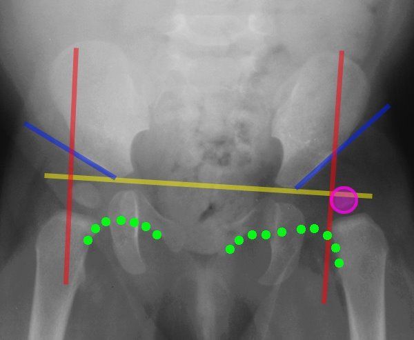
Lines Of The Hip Pediatrics Pediatrics Pediatric Nurse Practitioner Pediatric Radiology

Developmental Dysplasia Of The Hip Ddh Diagnostic Imaging Developmental Dysplasia Of The Hip Diagnostic Imaging Case Study